Printable Eye Anatomy Diagram
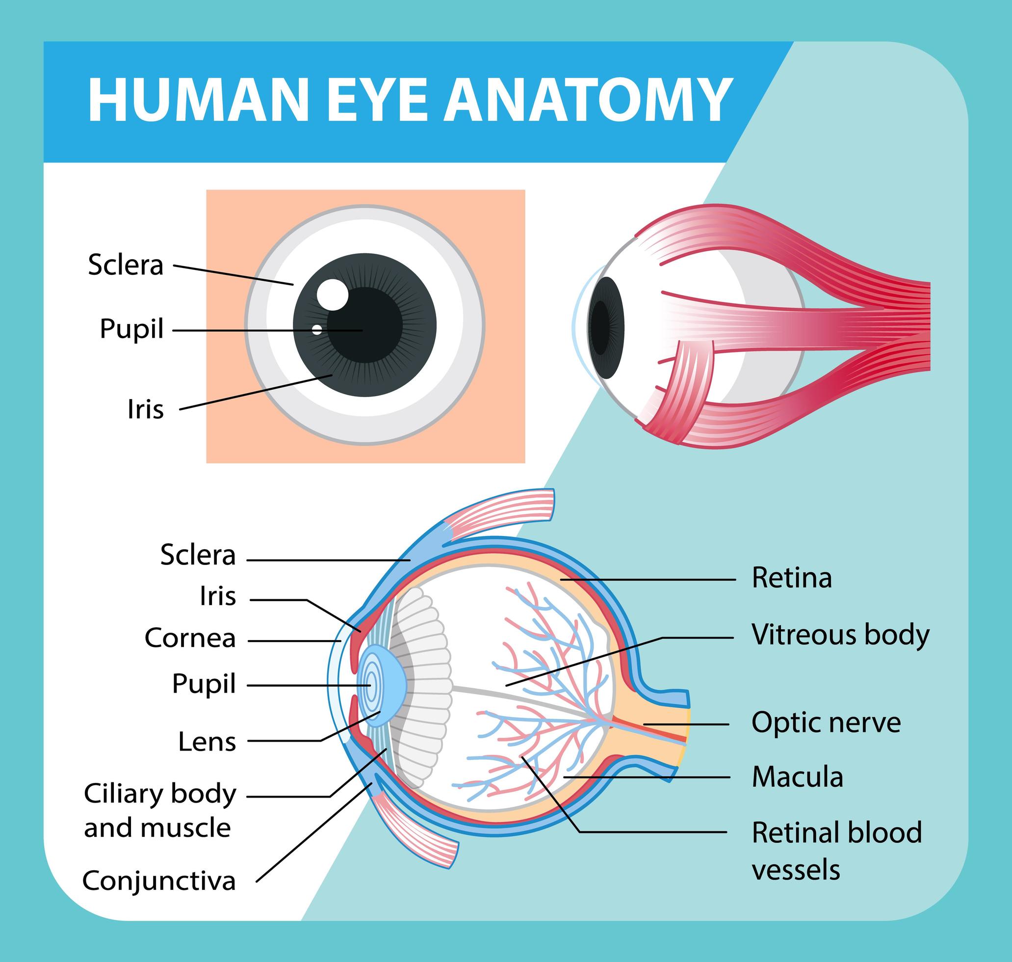
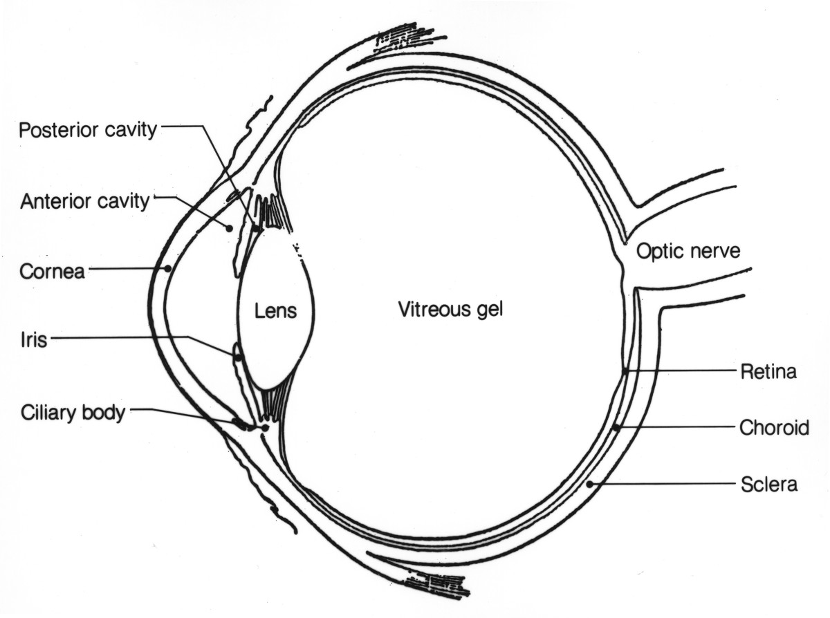
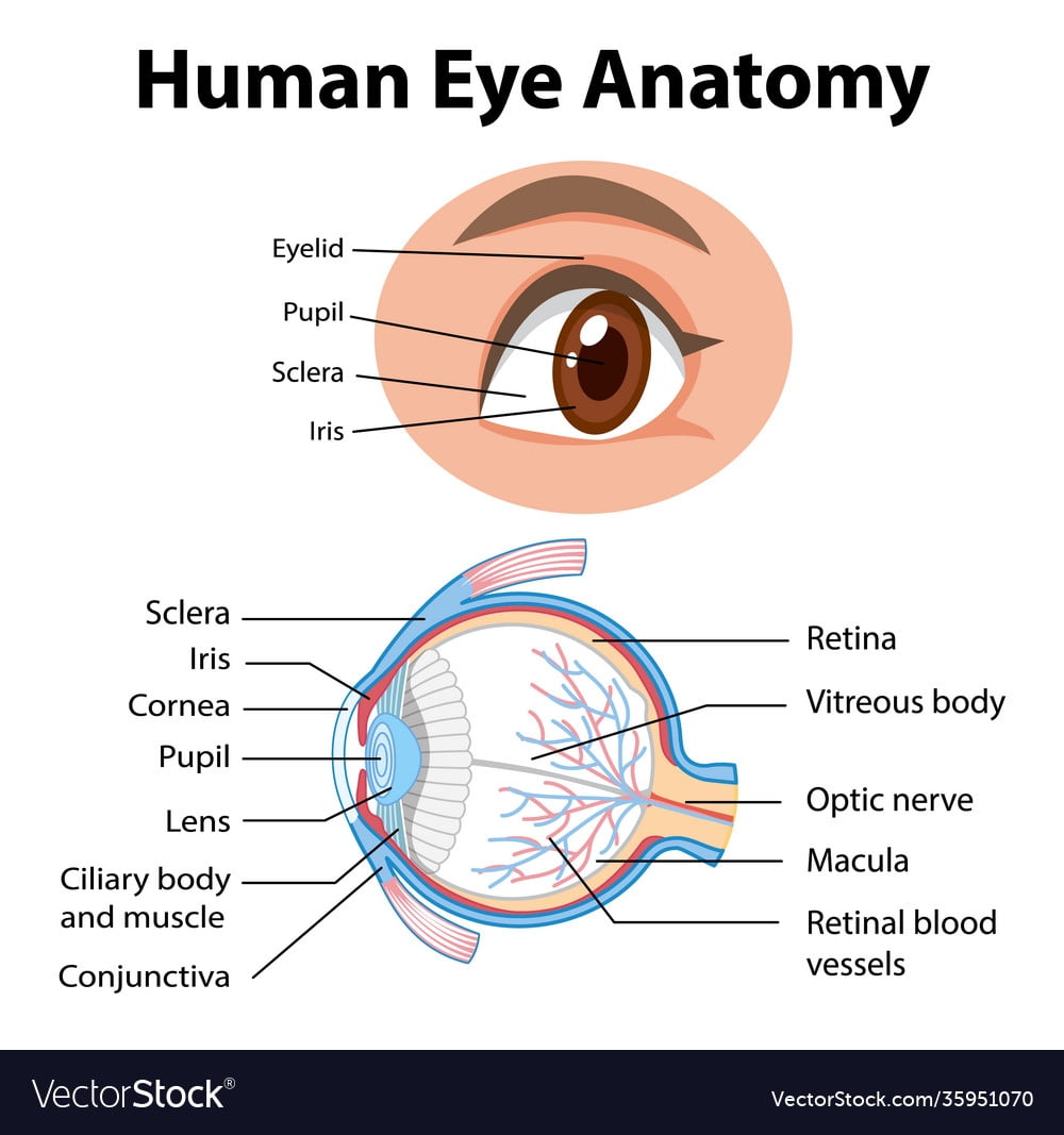
Printable Eye Anatomy Diagram - Some of them have labels to make it very easy to recognize parts of the body. A diagram to learn about the parts of the eye and what they do. The iris is the colored part of the eye that regulates the amount of light entering the eye. Web the eye has several major components: Amount of light entering the eye. The diagrams show cross sections of the human eyeball; The cornea is the clear outer part of the eye’s focusing system located at the front of the eye. Read an overview of general eye anatomy to learn how the parts of the eye work together. Anatomy of the eye anterior chamber lens pupil suspensory ligament word bank papilla viterous body lateral rectus muscle iris retina fovea conjunctiva choroid optic nerve aqueous humor ciliary body cornea medial rectus muscle posterior chamber sclera sc i encenotes. Web this printable contains 13 clear and simple cross sectional diagrams of the human eye.
Web diabetes and healthy eyes. Web in this interactive, you can label parts of the human eye. Eyes are approximately one inch in diameter. The iris is the colored part of the eye that regulates the amount of light entering the eye. The cornea is the clear outer part of the eye’s focusing system located at the front of the eye. Web eye anatomy (16 parts of the eye & what they do) the following are parts of the human eyes and their functions: Web 6 min read. Web get free human anatomy worksheets and study guides to download and print. Read an article about how vision works. As we journey through the different parts, refer to them to better understand their functions.
Web for a fun way to reinforce learning regarding the human body, you can use free printable eye anatomy worksheets. Here are descriptions of some of the main parts of the eye: Clear and flexible, the lens changes shape to focus light on the retina. The white visible portion of the eyeball. The iris is the colored part of the eye that regulates the. The iris is the colored part of the eye that regulates the amount of light entering the eye. From the bones of the orbit, to the eyeball and everything in between, you’ll be able to find the perfect quiz to help you tackle the seemingly scary topic of eye anatomy. Anatomy of the eye anterior chamber lens pupil suspensory ligament word bank papilla viterous body lateral rectus muscle iris retina fovea conjunctiva choroid optic nerve aqueous humor ciliary body cornea medial rectus muscle posterior chamber sclera sc i encenotes. Find out why the human eye has been called the most complex organ in our body. Web to understand the diseases and conditions that can affect the eye, it helps to understand basic eye anatomy.

eye diagram Discovery Eye Foundation
The lens is a clear part of the eye behind the iris that helps Web this article explores the anatomy of the human eye, looking at the different structures and their functions. Here is a tour of the eye starting from the outside, going in through the front and working to the back. As we journey through the different parts,.
Anatomy Of The Eye Diagram Labeled
Here is a tour of the eye starting from the outside, going in through the front and working to the back. Read an article about how vision works. The diagrams show cross sections of the human eyeball; Your eye is a slightly asymmetrical globe, about an inch in diameter. A diagram to learn about the parts of the eye and.

Free and Printable Eye Diagram 101 Diagrams
The lens is a clear part of the eye behind the iris that helps Clear and flexible, the lens changes shape to focus light on the retina. Web become a master of the eye anatomy with this specially designed quiz which covers bones and muscles of the orbit and eye anatomy (including neurovasculature)! Web we offer basic identification, advanced identification.

FileSchematic diagram of the human eye en.svg Wikipedia, the free
Web anatomy of the eye. Check out our top picks below! The lens is a clear part of the eye behind the iris that helps The iris is the colored part of the eye that regulates the amount of light entering the eye. Anatomical areas the quadrangular space.

Diagrams of the Human Eye 101 Diagrams
Web for a fun way to reinforce learning regarding the human body, you can use free printable eye anatomy worksheets. Muscles of the leg muscles in the anterior compartment of the leg. The cornea is the clear outer part of the eye’s focusing system located at the front of the eye. Web anatomy of the eye. Drag and drop the.

Schematic Diagram Of Human Eye
Web here are descriptions of some of the main parts of the eye: The conjunctiva is the membrane covering the sclera (white portion of your eye). They photocopy well and are great for use as a labeling and coloring exercise for your students. Carries the messages from the retina to the brain. A diagram to learn about the parts of.
Anatomy of the Eye Human Eye Anatomy Owlcation
Eyes detect both brightness and color. From the bones of the orbit, to the eyeball and everything in between, you’ll be able to find the perfect quiz to help you tackle the seemingly scary topic of eye anatomy. Web become a master of the eye anatomy with this specially designed quiz which covers bones and muscles of the orbit and.

Printable Human Eye Diagrams 101 Diagrams
These are pdf, png, and google slides worksheets. Web for a fun way to reinforce learning regarding the human body, you can use free printable eye anatomy worksheets. The conjunctiva is the membrane covering the sclera (white portion of your eye). Your eye is a slightly asymmetrical globe, about an inch in diameter. Use your mouse or finger to hover.

Eye Diagram Vector Art, Icons, and Graphics for Free Download
Web diabetes and healthy eyes. Eyes are approximately one inch in diameter. Web become a master of the eye anatomy with this specially designed quiz which covers bones and muscles of the orbit and eye anatomy (including neurovasculature)! The diagrams show cross sections of the human eyeball; Web in this interactive, you can label parts of the human eye.
Printable Eye Anatomy Diagram Printable Lab
Web for a fun way to reinforce learning regarding the human body, you can use free printable eye anatomy worksheets. Use your mouse or finger to hover over a box to highlight the part to be named. Web here are descriptions of some of the main parts of the eye: Web our eyes are organs that let us see. Human,.
Here Is A Tour Of The Eye Starting From The Outside, Going In Through The Front And Working To The Back.
The iris is the colored part of the eye that regulates the amount of light entering the eye. The conjunctiva is the membrane covering the sclera (white portion of your eye). Eyes are approximately one inch in diameter. Your eye is a slightly asymmetrical globe, about an inch in diameter.
The Front Part (What You See In The Mirror) Includes:
The cornea, pupil, lens, iris, retina, and sclera. Web anatomy of the eye. Eyes detect both brightness and color. Web our eyes are organs that let us see.
The Iris Is The Colored Part Of The Eye That Regulates The Amount Of Light Entering The Eye.
System located at the front of the eye. The diagrams show cross sections of the human eyeball; Web for a fun way to reinforce learning regarding the human body, you can use free printable eye anatomy worksheets. The cornea is the clear outer part of the eye’s focusing system located at the front of the eye.
The Cornea Is The Clear Outer Part Of The Eye’s Focusing.
The white visible portion of the eyeball. Here are descriptions of some of the main parts of the eye: The lens is a clear part of the eye behind the iris that helps Web to understand the diseases and conditions that can affect the eye, it helps to understand basic eye anatomy.